© elfruler 2018, 2026
with thanks to Donna Young
(Click on images for an enlarged view; on videos, click on full screen icon. )
The avian egg is a marvel of nature, a self-enclosed and perfectly effective living environment for the developing bird embryo. It contains all that is necessary to enable a small and weak organism to develop into a chick with all its parts and enough strength to break through and emerge into the outside world. Here is an account of the many factors involved in a chick’s hatching.
Inside the shell
- The eggshell is a complex structure of hard calcium carbonate crystals interwoven with collagen fibers, and it is coated by a thin layer of crystalline calcite and smooth protein cuticle. The shell is sturdy enough to protect the developing embryo, yet it has microscopic pores that allow oxygen to pass into the egg and carbon dioxide and water vapors to pass out.

- Two soft keratin membranes line the inside of the shell, both formed in the isthmus of the oviduct to serve as developmental and structural foundation for the hard shell. These shell membranes help protect the embryo from external microbes, facilitate the exchange of oxygen, carbon dioxide, water between the embryo and the outside environment, and collect calcium from the shell to be transferred to the embryo for its bone development. The outer membrane is attached to the inside of the shell, and the thinner inner membrane lines the outer membrane. There is a small gap between the 2 membranes in the blunt end of the shell.
- The yolk sac, with its supply of fats and proteins to nourish the growing embryo, is attached to the embryo by a cord leading into the abdominal cavity.
- Another membrane is adjacent to the inner shell membrane, the chorioallantoic membrane (CAM). It surrounds the embryo and the yolk sac and is connected to the embryo’s heart via blood capillaries that pass through the abdominal wall at the same opening where the yolk sac enters. The CAM performs the embryo’s respiratory function, transferring oxygen received by the shell membranes to the embryo and sending carbon dioxide out through the shell membranes. It also transfers calcium from the shell to the embryo, and collects wastes and stores them until after hatch. At hatch the CAM remains attached to the shell and detaches from the chick.
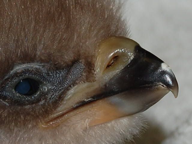
- Starting about a third of the way through the ~36 days of an eagle embryo’s growth in the egg, an “egg tooth” or “pipping tooth,” a small, hard, sharp protuberance of calcified keratin near the tip of the beak’s upper mandible, begins to develop. Click here for a closeup of the egg tooth on a hatchling eaglet at the Institute for Wildlife Studies. The egg tooth gradually wears away within a couple of weeks after hatch.
- A few days before hatch begins, a paired muscle in the back of the chick’s neck (the complexus or hatching muscle) swells in response to the influx of fluids from adjacent lymph glands. (This muscle plays a role in neck extension in grown eagles).
- Just before hatch, the egg weighs about three-quarters of its weight when it was laid, because it has absorbed and metabolized fats from the yolk and lost evaporated water through the membranes. When laid, a Bald Eagle weighs about 113-127 g (4-4.5 oz.). By the time it hatches the egg’s weight has been reduced to about 89-102 g (3.2-3.6 oz.). Sizes vary with latitude, larger in the north than in the south, and with hatch order – eggs decrease in size from the first egg of a clutch to the last. The eggshell is much thinner than when the egg was laid because the chick has absorbed much of its calcium for its bones.
The hatching process
- As the embryo nears hatch it takes up most of the space inside the shell – it is crowded in there! The chick lies on its left side with its feet in the smaller end of the shell and its head tucked forward against its breast near the blunt end of the shell. The inner shell membrane and CAM are draped over the head, beak, and wings.

© elfruler - In the days leading up to hatch, the evaporation of water leads to an increase in the amount of air inside the shell, which enlarges the gap between the inner and outer shell membranes at the blunt end. This expanded air cell spreads along the shell’s upper side. As it takes hatching position the embryo’s back is pressed against the air cell and its head is tucked forward towards its belly and under the right wing, with the beak pointing toward the air cell.

Image used by permission courtesy of Charles County, MD, Dept. of Recreation, Parks, & Tourism, Port Tobacco River Park Eagle Cam - The embryo absorbs the remainder of the yolk sac into its abdominal cavity and the opening closes up, leaving a “yolk sac scar” (the umbilicus or “belly button”), which eventually fades. The protruding umbilicus is obvious in this screenshot from the MD Port Tobacco cam in 2019.
- The swollen complexus muscle in the back of the neck contracts, causing the body of the embryo to stretch out and pulling the head back so that it presses against the inner shell membrane. The right wing further stretches the membrane and lifts it away from the head. The pressure from head, beak, and wing ruptures the membrane, resulting in what is called the internal pip. The air cell releases a small supply of oxygen and prompts the chick’s lungs and its 9 air sacs to begin functioning.
- With its lungs beginning to work, the chick (as it now can be called, instead of embryo) begins to emit little cheeps, which often can be heard on the nest cams.
- After the internal pip the chick rests as its lungs begin to inhale oxygen and exhale carbon dioxide and to make the proper exchange with the circulatory system. The respiratory function of the CAM is winding down and the cord connecting it to the embryo’s heart through the umbilicus site begins to dry up.
- While the lung function is developing, the contractions of the complexus muscle increase. The head and beak begin to jerk back against the shell repeatedly and the spine and legs push against the shell, finally piercing it with the egg tooth, ideally about ¼-⅓ of the way down the side of the shell near the blunt end of the egg. (If the pip is nearer the smaller end of the shell, the chick’s body probably is not positioned properly and it may have more difficulty hatching.) This is seen from the outside as a tiny hole, called the external pip. This 1-minute video offers a rare view of the beginning of an external pip, at the Decorah nest in 2023. (Video used by permission under Creative Commons license, courtesy of Raptor Resource Project.)
(The actual time elapsed was 6 minutes, I have sped it up x 6.)

Image used by permission courtesy of ND-LEEF Eagle Cam - The external pip may take a star-like appearance (“starring”), as in this screenshot from the IN Notre Dame cam in 2021. On the nest cams, the beginning of the external pip may not be in view, hidden by an incubating parent or nest materials, or because it is turned away from the cam.
- The external pip allows abundant outside oxygen directly into the egg. The chick rests again at intervals while its respiratory and circulatory systems continue to adapt. When the incubating parent rises from the nest cup, if the chick is awake, its cheeping can be heard. Here is video of a hatching egg at the CO Fort St. Vrain nest in 2021, with the cheeping quite audible. (Video used by permission under Creative Commons license, courtesy of Xcel Energy Fort St. Vrain Station, Colorado & Raptor Resource Project.)
- The external pip accelerates fluid loss from the egg and the chick’s body, which can be good because a slightly reduced body mass allows the chick more room to maneuver as it pushes against the shell. However, if the ambient humidity is low, the exposed shell membranes can dry up, and their leathery texture may hold the shell together and can be harder for the chick to tear apart, making hatching more difficult.
- The initial pip increases in size over the next few hours. In the early stages, small bits of shell might bulge from the hole, sometimes visible in profile if the egg is turned sideways to the cam. The egg tooth pokes and scrapes the shell, and the legs, shoulders, neck, and back flex, breaking up the shell and creating larger holes and cracks. The chick’s beak, head, a wing, or a foot might poke through. The enlarged complexus muscle at the nape provides cushioning and support during this process. The chick rests between efforts.
- As its legs flex and contract, the chick may rotate inside the shell counterclockwise, possibly turning a third or halfway or more round the shell as hatching continues. This rotation can result in a roundish disc at the blunt end of the shell, a “cap,” which pops off and exposes the chick’s head and wings. Researchers call this “symmetrical hatching,” referring to the more or less symmetry between the two parts of the shell. I call this a “clean” hatch.
 Here is a screenshot of the chick emerging out of the egg at the BC Boundary Bay Central nest cam in 2024, with its feet still in the larger section at the pointed end, its head out of the smaller cap and curled under between its wings. (Image used by permission under Creative Commons license, courtesy of Hancock Wildlife Foundation.) And here is video of the final stage of a symmetrical hatch in 2025 at the National Conservation Training Center nest in WV. (Video used by permission courtesy of U.S. Fish & Wildlife Service, National Conservation Training Center.)
 - While symmetrical hatching is the norm for most avian species, observers of Bald Eagle cams have noted that not all hatches result in a clean breakup of the shell. Sometimes the first external pip seems to simply grow in size until the chick breaks through the gap. Occasionally the shell membranes hold the shell together so that it does not break apart cleanly. I call this a “messy” hatch, which in most cases ends successfully, even if it takes a bit longer than a symmetrical hatch. This happened to both eggs at the White Rock nest in British Columbia in 2025. (Image used by permission under Creative Commons license, courtesy of Hancock Wildlife Foundation.)
 Note the wrinkled shell membrane underlying the shell in the egg on the right (the first laid). The time from first sighting of the first egg’s pip to its hatch was 42 hours 17 minutes, 35 hours 5 minutes for the second egg. Both eaglets hatched successfully and fledged. The first egg holds the record in my stats for the longest time between oviposition and hatch: 42 days 10 hours 50 minutes. (A member of the Hancock Wildlife Forum documented the hatches of these eggs in this remarkable series of videos.)
 - Many biologists and observers consider the egg to be “hatched” when the shell is broken apart and the chick is free. But “free” can be open to interpretation. The strictest definition of a completed hatch is that the chick is lying completely separate from any of the shell. Less strictly, “free” could mean that its head, feet, or rump are lying on or in part of the shell but not covered or restricted by it. Some viewers take the view that the chick is hatched even if the cap is atop the head like a helmet, or the feet or rump are inside the smaller end of the shell like a sleeping bag or a diaper. Others point out that once the shell is broken in two, it not going to be put together again (reference Humpty Dumpty), and the chick is hatched even if it hasn’t fully emerged. Often the cams do not provide a full view of the hatch, however “hatch” is interpreted. At some cams the “official” hatch time is defined as the moment that the cam view finally reveals the chick’s entire body away from the shell.
- The new hatchling is covered with a thin layer of downy feathers – its natal down – which is damp from the fluids inside the shell, and its skin is mostly pinkish (but dark gray around the eyes). The down will dry out within a short time (it may even start drying out when the shell begins to break up), and the chick is then covered with a soft, fluffy, light gray coat.
- The hatching process is strenuous and can take a couple of days or more or more to complete. The new hatchling rests for a while, but soon it can become fairly active, although its movements are awkward and erratic – stretching and flexing, rolling around, lifting its head or a wing, cracking open its eyes briefly, occasionally cheeping. The complexus muscle recedes in size and the chick’s neck isn’t yet strong enough to hold its head up.
Parental behavior during hatching
- Starting several days before hatching begins, both parents – especially the male – may bring food to the nest in anticipation of both the chick’s and the mother’s need for food as brooding begins.
- The parents are aware that the hatch has begun when they hear the chick’s vocalizations after the internal pip and possibly also hear its pecking and scraping at the shell, even before the external pip. The incubating adult may stand above or to the side of the egg and lean in or cock its head, seeming to listen. Parents may chirp softly to the chick. Click here for video of an incubating parent hearing its chick cheeping before the external pip at the USFWS National Conservation Training Center nest in WV.
- They may exhibit restlessness in the egg cup, rising and sometimes circling the cup every few minutes to check the eggs.
- They might gently nudge the hatching egg and the emerging chick. They may push shell fragments away from the hatching egg.
- They often pull soft nesting material in toward the nest cup (sometimes building a wall between the cup and the viewers!).
- The parents by instinct do not assist the chick in breaking the shell. They take great care not to pick at or unduly jostle the hatching egg. Active participation in breaking apart the shell could damage the still fragile blood vessels in the CAM. The arduous effort of hatching is the chick’s first physical workout, important preparation for life outside the shell.
- We are spoiled by the nest cams that give us views that provide more information than was possible before, although some of the cams do not give a clear view into the nest cup so that we can watch eggs hatch. But even if we can’t see the hatch itself, parental behavior can provide clues that it is eminent or occurring, such as preparatory provision of food, listening for cheeps from an egg, restless upping, downing, circling, and nudging the eggs every few minutes, and most obviously, once a hatchling is ready, offering it food.
Post-hatch
- Bald Eagle hatchlings are “semialtricial,” which means they are nearly helpless when they hatch, with limited motor skills and strength, entirely dependent on parents for food and warmth, and confined to the nest (“nidicolous” – “nest inhabiting”). All raptors are semialtricial and must spend several weeks being cared for by their parents before they fledge and are capable of fending for themselves.
- Bald Eagles are not considered fully altricial (like songbirds and parrots) because their eyes are open at hatch, they are covered with downy feathers, and they have some mobility.
- At the other end of the developmental spectrum from altricial are “precocial” chicks, like geese, ducks, swans, chickens, and quail, which are capable of walking (and often swimming) and thermoregulating soon after they hatch. They are “nidifugous” (“nest fleeing”) meaning they can leave the nest almost immediately after hatching.
- In the days before it hatched the chick has absorbed the yolk sac into its body, whose nutrients feed it in the few hours before and after hatch. It will not need to be fed by its parents for several hours.
Clearly, hatching is a complex process, and most of the time it ends successfully. Sometimes, though, things can go wrong. This page surveys reasons why an egg might fail to hatch.
A rich description of how a bird egg hatches is excerpted from Tim Birdhead’s book The Most Perfect Thing: Inside (and Outside) a Bird’s Egg (2016) on The Audubon Society website
Here is a compilation video of the hatch of the first eaglet at the West End nest on Catalina Island on 20 March 2018.
Dramatization of the development of a chicken embryo from oviposition to hatch (21 days)
References
- Bond, G. M., V.D. Scott, and R. G. Board 1986. Correlation of mechanical properties of avian eggshells with hatching strategies. Proceedings of the Zoological Society of London (A)209:225-237.
- Bond, G. M., R. G. Board,and V. D. Scott 1988. An account of the hatching strategies of birds. Biological Review63: 395-415.
- Bortolotti, Gary R. 1984. Physical development of nestling Bald Eagles with emphasis on timing of growth events. Wilson Bulletin96: 524-542.
- Butcher, G.D., and N.A. H. 2002. Chicken Embryo Malpositions and Deformities. University of Florida Institute of Food and Agricultural Sciences (IFAS)
- Cobb Hatchery Management Guide. 2020. Cobb Genetics. (esp. pp. 78 and 81) https://www.cobbgenetics.com/resources/management-guides
- Deeming, D. C. 2002. Avian Incubation: Behaviour, Environment, and Evolution(Oxford and New York: Oxford University Press).
- Deeming, D. C. and S. J. Reynolds, eds. 2015. Nests, Eggs, and Incubation: New Ideas about Avian Reproduction(Oxford: Oxford University Press).
- Drent, Rudolf 1973. The natural history of incubation. In Breeding Biology of Birds: Proceedings of a symposium on breeding behavior and reproductive physiology in birds, Denver, Colorado, February 1972, ed. Donald S. Farner (Washington, DC: National Academy of Sciences): 262-322.
- Fox, Nick 1995. Understanding the Bird of Prey(Surrey, British Columbia and Blaine, WA: Hancock House Publishers).
- Gross, G.H. 1985. Innervation of the Complexus (“Hatching”) Muscle of the Chick. Journal of Comparative Neurology 232: 180–189.
- Gill, Frank B. 2007. Ornithology, 3rd ed. (New York: W. H. Freeman and Company).
- Hamburger, Viktor and Ronald Oppenheim 1967. Prehatching motility and hatching behavior in the chick. Journal of Exp. Zool. 166: 171-204
- Lovette, Irby J. and John W. Fitzpatrick, eds. 2016. The Cornell Lab of Ornithology Handbook of Bird Biology, 3rd ed. (Chichester, West Sussex: John Wiley & Sons, Ltd.
- Oppenheim, Ronald W. 1972. Prehatching and hatching behaviour in birds: a comparative study of altricial and precocial species. Animal Behaviour20: 644-655.
- Podulka, Sandy, Ronald W. Rohrbaugh, Jr., & Rick Bonney, eds. 2004. Handbook of Bird Biology, 2nd ed. (Ithaca, NY: The Cornell Lab of Ornithology).
- Proctor, Noble S. and Patrick J. Lynch 1993. Manual of Ornithology: Avian Structure & Function(New Haven and London: Yale University Press).
- Sharpe, Peter 1995. Guide to Bald Eagle Egg Incubation and Chick-Rearing. Institute for Wildlife Studies.
- Starck, J. Matthias and Robert E. Ricklefs, eds. 1998. Avian Growth and Development Evolution within the Altricial-Precocial Spectrum(New York and Oxford: Oxford University Press).
- Starck, J.M. 2021. Morphology of the avian yolk sac. Journal of Morphology 282: 959–972.
- Tullett, S. 2009. Investigating Hatchery Practice. (esp. Pp. 14-15) https://aviagen.com/assets/Tech_Center/Ross_Tech_Articles/RossTechInvestigatingHatcheryPractice.pdf.
You must be logged in to post a comment.